Our profession is dedicated to providing the best care to each patient individually and by using advanced technology and scientifically proven treatment methods it improves oral health. Radiography can help a dentist diagnose many oral diseases. It is left to the dentist to assess the benefit of making an X-ray compared to the risk of excessive radiation and the cumulative effect of radiation over time. The dentist, familiar with the history of the patient's disease, should evaluate which type of X-ray imaging is best suited for treating the given condition. In order for our patients not to have any dilemmas about the type and frequency of radiation in dental practice, we will provide all necessary information from the official site of the International Atomic Energy Agency (IAEA) as well as guidelines for radiography given by the American Dental Association.
What is meant by "radiation dose"?
Radiation dose is the amount of energy absorbed during exposure to X rays. This dose is important as the absorbed energy can cause biological damage to the person who has been exposed to radiation. There are two ways to determine the radiation dose in dentistry: the estimated dose on the patient's skin surface (air kerma) and the effective dose. The dose on the skin surface is easier to measure while the calculation of the effective dose is more complicated but the obtained value can be associated with the risk of radiation.
Which units are used to measure the dose of X-radiation?
The unit for air kerma is Gray (Gy), but in dentistry, the radiation doses used are usually a small fraction of one grey (miligray - mGy, or even microgray μGy). The unit for effective dose is Sievert (Sv). In dental radiology, a dose consists only of fractions of a single sievert (milisievert, mSv or microsievert μSv).
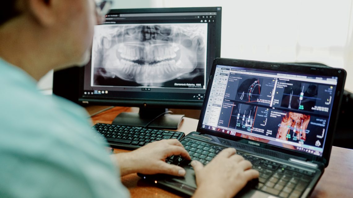
The following doses are common for different types dental images:
- 1-8 mGy for intraoral images
- about 100 mGy for panoramic images (orthopantomograph)
- 0.25-7 mGy for cephalometric images
Effective doses are:
- 1-8 μSv for intraoral images
- 4-30 μSv for panoramic images (orthopantomograph)
- 2-3 μSv for cephalometric images
- 34-652 μSv when imaging smaller dentoalveolar regions by computerized tomography (CBCT) and 30-1079 μSv when imaging large craniofacial regions
Radiation doses during intraoral and cephalometric radiography are generally lower than daily natural radiation. In panoramic imaging, the values vary, but even the highest possible values are equivalent to those in a chest x-ray or doses of several days of natural radiation. CBCT has a wide range of values and can be tens or hundreds of μSv of effective dose more than with conventional radiographic techniques. In view of this, the American Dental Association has adopted a declaration on the use of CBCT:
"As with other radiographic modalities, CBCT imaging should be used only after a review of the patient's health and imaging history and the completion of a thorough clinical examination. Dental practitioners should prescribe CBCT imaging only when they expect that the diagnostic yield will benefit patient care, enhance patient safety or improve clinical outcomes significantly."
Radiographic imaging of pregnant women
The patient is to deliver information on possible pregnancy to the dentist. For each woman in the reproductive period, pregnancy should be suspected until proven otherwise. If the patient is pregnant, all necessary information should be collected by reviewing the patient’s health history and through a thorough clinical examination. If radiography is necessary, the pregnant woman should be informed of the potential risk of radiation to the fetus.
If the X-ray is made, what are the risks to the fetus?
The radiation of several μSv is extremely low, and therefore the risk to the fetus is low. The risk of developing cancer in a still unborn child at 10 μSv radiation is several thousand times lower than the risk of developing cancer if there is a positive family history. The risk of genetic anomalies due to radiation is far lower than with the genetic predisposition to anomalies. Therefore, radiation doses in everyday dental practice should not instigate thinking about terminating pregnancy and the patient with these dilemmas should be adequately advised.
Should the dentition in children be viewed using a panoramic image?
No, this procedure is not justified as a routine one. Radiography should be used when a clinical examination suspects of the presence of anomalies or when planning an orthodontic therapy.
Source:
https://www.iaea.org/resources/rpop/health-professionals/dentistry/radiation-doses
https://www.ada.org/~/media/ADA/Member%20Center/FIles/Dental_Radiographic_Examinations_2012.pdf